The Of Treatments for Premature Ejaculation: What Works Best?

The Greatest Guide To Oral Rivaroxaban for the Treatment of Symptomatic
Blood tests also can determine the quantity of oxygen and carbon dioxide in your blood. An embolism in a blood vessel in your lungs may lower the level of oxygen in your blood. In addition, blood tests may be done to figure out whether you have actually an inherited clotting condition. Chest X-ray, This noninvasive test shows pictures of your heart and lungs on film.

Current Endovascular Treatment Options in Acute Pulmonary Embolism - Journal of Clinical Imaging Science
Ultrasound, A noninvasive test understood as duplex ultrasonography (often called duplex scan or compression ultrasonography) uses sound waves to scan the veins in your thigh, knee and calf, and in some cases in your arms, to look for deep vein embolism. A wand-shaped device called a transducer is moved over the skin, directing the sound waves to the veins being evaluated.

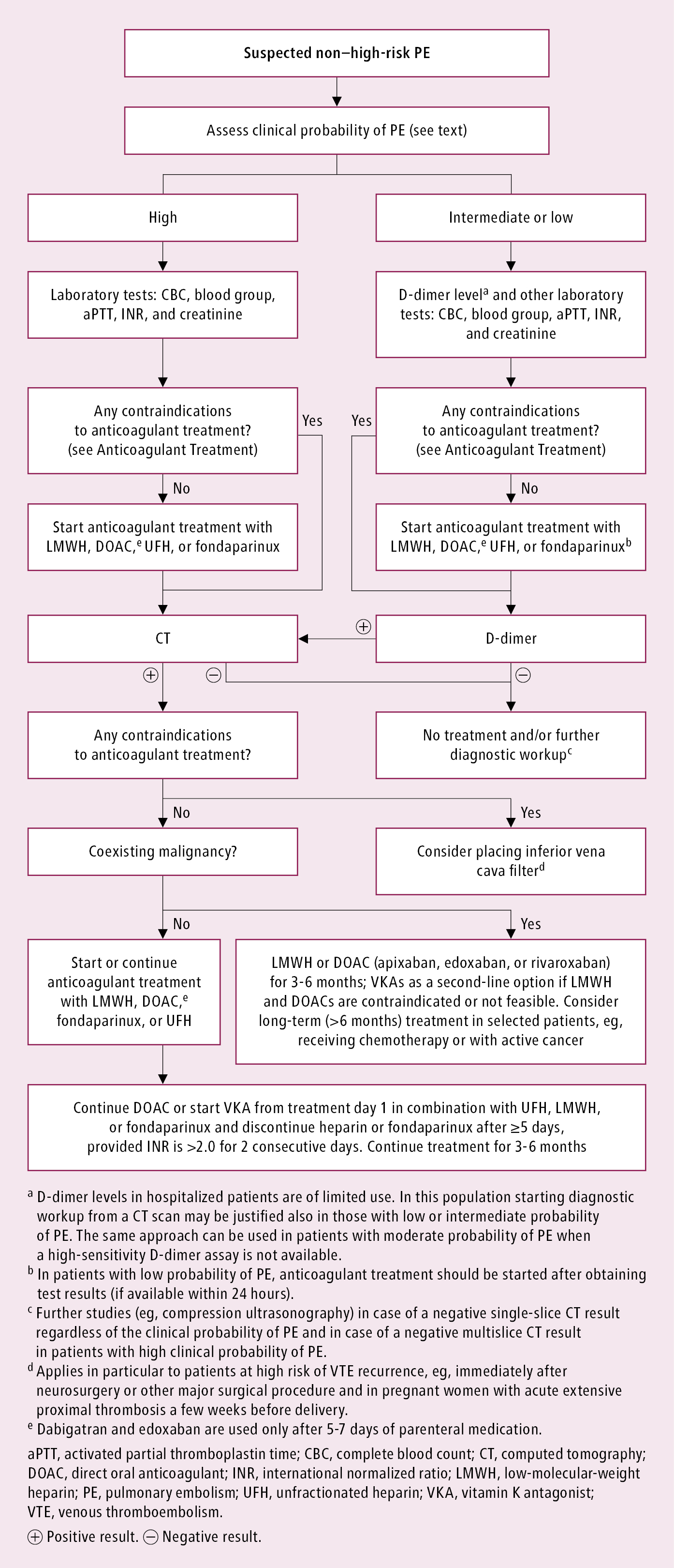
PE treatment based on early mortality risk- Download Scientific Diagram
The absence of embolisms reduces the possibility of deep vein thrombosis. If clots are present, treatment likely will be started instantly. CT lung angiography, CT scanning produces X-rays to produce cross-sectional images of your body. Check For Updates called CT lung embolism research study creates 3D images that can detect irregularities such as pulmonary embolism within the arteries in your lungs.

Some Known Facts About Pulmonary Embolism (Blood Clot in Lung) Symptoms, Causes.
Ventilation-perfusion scan (V/Q scan)When there is a requirement to prevent radiation direct exposure or contrast from a CT scan due to a medical condition, a V/Q scan may be performed. In this test, a tracer is injected into a vein in your arm. The tracer maps blood circulation (perfusion) and compares it with the air flow to your lungs (ventilation) and can be utilized to identify whether blood clots are causing signs of lung high blood pressure.

Developing a complex intervention for the outpatient management of incidentally diagnosed pulmonary embolism in cancer patients - BMC Health Services Research - Full Text
It's the most accurate way to diagnose pulmonary embolism, but due to the fact that it requires a high degree of skill to administer and has possibly serious threats, it's typically carried out when other tests stop working to provide a definitive medical diagnosis. In a lung angiogram, a versatile tube (catheter) is inserted into a big vein usually in your groin and threaded through your heart and into the lung arteries.
In some individuals, this treatment might trigger a temporary modification in heart rhythm. In addition, the color might trigger increased risk of kidney damage in individuals with minimized kidney function. MRIMRI is a medical imaging technique that uses a magnetic field and computer-generated radio waves to develop detailed pictures of the organs and tissues in your body.
